Chinese
 English
|
|
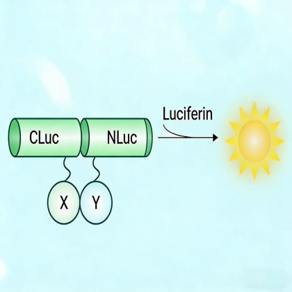
Service Introduction Luciferase complementation imaging detection refers to the method used to determine whether two target proteins interact. The luciferase enzyme is divided into two parts: the N-terminal (NLuc) fragment 2–416 and the C-terminal (CLuc) fragment 398–550, which serve as reporter proteins. The genes encoding the N-terminal (NLuc) fragment and the C-terminal (CLuc) fragment are constructed into different expression vectors to obtain N-terminal and C-terminal vectors. In the N-terminal vector, the N-terminal (NLuc) fragment's amino terminus is fused with the gene of one target protein to create the N-terminal detection vector, while in the C-terminal vector, the C-terminal (CLuc) fragment's carboxyl terminus is fused with the gene of another target protein to create the C-terminal detection vector. When the expression products of the N-terminal and C-terminal detection vectors are mixed, the two parts of the reporter proteins downstream of the target proteins do not spontaneously assemble. Only when the target proteins fused to these two reporter fragments interact can the two reporter proteins correctly assemble and generate activity. After adding a luminescent substrate, the chemiluminescence can be detected using specific instruments. The luminescence is then used to determine whether the two target proteins interact. Service Advantages ▶High sensitivity: LCA can detect protein interactions at levels as low as picomoles, making it highly suitable for studying interactions of low-abundance proteins. ▶Real-time monitoring: Because the luciferase-catalyzed reaction is rapid and the bioluminescent signal is strong, LCA enables real-time monitoring of protein interactions without the need to fix or lyse cells, thus reducing loss and variability during sample processing. ▶Non-invasive detection: LCA is conducted within live cells without disrupting cell structures, allowing observation of protein interactions under physiological conditions and providing interaction information in a more realistic biological environment. ▶Wide dynamic range: LCA can cover a broad range of interaction strengths, detecting interactions from weak to strong. Service Process
Article Case A flavonoid metabolon: cytochrome b5 enhances B-ring trihydroxylated flavan-3-ols synthesis in tea plants academic journal:The Plant Journal
Split-luciferase complementation assay analyses the spatial proximity between CsC4H, CsF30 H, and CsF30 50 H proteins in N. benthamiana tobacco leaves. N.C., negative control, inject empty vector pC1300-nLUC and pC1300-cLUC together; N.C.1–3, negative control, inject empty vector pC1300-cLUC and P450-nLUC together; N.C.4–6, negative control, inject empty vector pC1300-nLUC and cLUC-P450 together. Applicable Fields ▶Protein interaction studies: LCA can monitor direct or indirect interactions between proteins in living cells in real time, providing data for understanding cellular signal transduction, complex assembly, and protein networks. ▶Plant science: LCA is applied in areas such as plant hormone responses and stress signaling, promoting crop improvement and agricultural production. ▶Signal transduction analysis: LCA helps reveal how signaling proteins interact and transmit signals within cells, offering in-depth insights into the molecular mechanisms of signal transduction pathways. ▶Pathogen-host interactions: LCA plays a role in elucidating pathogen infection mechanisms, host defense responses, and immune reactions, enhancing understanding of infectious diseases. |